In this article, our method to measure structure is used to non-invasively separate highly aggressive brain tumors from less aggressive ones.
Microstructure of tumor tissue can be examined non-invasively using MRI. However, it is not well-established how differences in MRI measurements are coupled to tissue microstructure. To investigate how these two are linked, the following hypothesis in brain tumors was tested: Variation in diffusion, caused by the size, shape, and orientation of cells, is associated with variations in tumor cell appearance and tumor cell density.
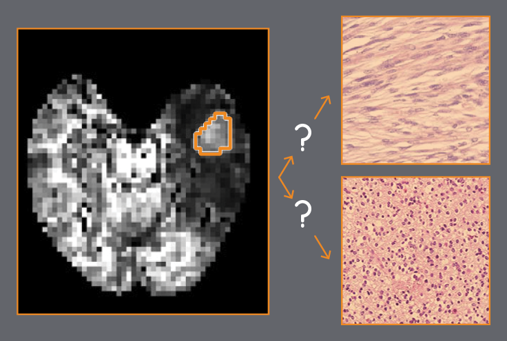
This was investigated by using our novel method to measure cell structure in 7 meningiomas (low aggressive brain tumor) and 8 gliomas (high aggressive brain tumor), before surgery. Results where correlated to parameters of tumor cell appearance and tumor cell density, examined by a pathologist. An excellent agreement between the parameters was observed, showing that our method allows non-invasive mapping of parameters that reflect variation in cell appearance and cell density. Thus, our method improves the interpretation of tumor microstructure, compared to current methods.